Art-labeling Activity: Structure of a Typical Synovial Joint
6 Osseous tissue IP. Its articulating surfaces movements stability and the clinical relevance.
Solved Art Labeling Activity Structure Of A Typical Chegg Com
No movement bursae reduce friction between adjacent structures but tendon sheaths do not.

. The costotransverse ligament fills the narrow interval between the vertebral transverse process and the rib neck. Art labeling activity -structure of a nail superficial and cross-sectional viewsjpg. The elbow is the joint connecting the upper arm to the forearm.
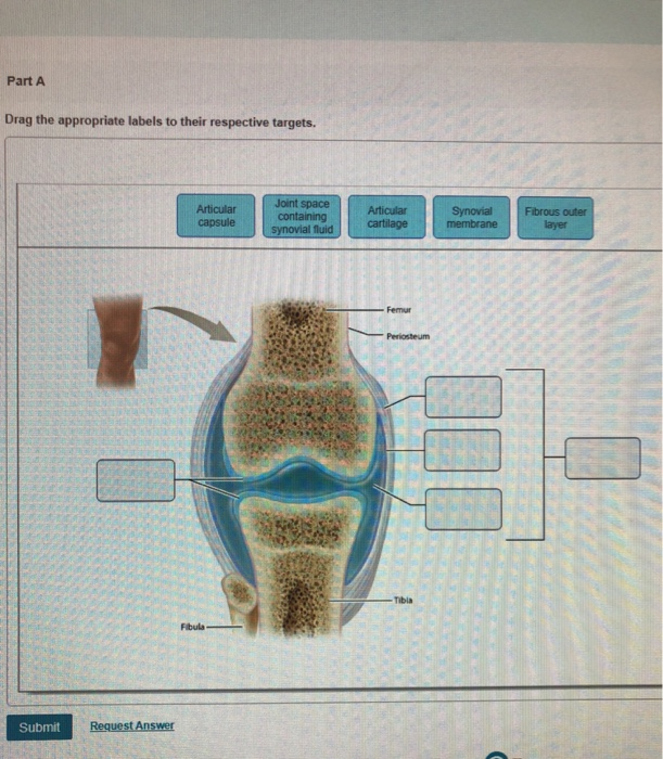
Types of Synovial Joints. In synovial joints the ends of. Label the diagram of a typical synovial joint using the terms provided in.
Muscles of the Chest Abdomen and Thigh Deep Dissection 1 of 2jpg. Synovial fluid is made by the synovial membrane. Reset Help Fibula Lateral meniscus Anterior cruciate ligament Ligaments That Stabilize the Knee Joint Tbial collateral ligament Articular cartilage Fibular collateral ligament Patellar surface Tibia Patelar igament cut Posterior cruciate ligament Medial meniscus.
The joints are reinforced by the costotransverse superior and lateral costotransverse and accessory ligaments. Learn vocabulary terms and more with flashcards games and other study tools. Synovial joints have synovial fluid in the joint cavity that lubricates or oils the joint so it moves smoothly.
Correct Spotlight Figure 82. Synovial Membrane inner- secretes produces synovial fluid for lubrication Synovial Membrane - makes the synovial fluid and encloses - a sheet of cells that lines the joint cavity Joint Cavity. Art labeling activity - ventricles of the brain Lateral viewjpg.
Fibrous Capsule outer- provides joint stability 2. Miami Dade College Miami SPC MISC. Circle the correct underlined term.
Skin structure Figure 48c. Multiaxial movement movement in or around three. Clinical Case Study.
Joint Motion Read through Spotlight Figure 82 and then complete the questions and activity below. A Structural Classification of Synovial Joints Label the types of synovial joints. A Pivot joints allow for rotation around an axis such as between the first and second cervical vertebrae which allows for side-to-side rotation of the head.
There are two types of cartilaginous joints. Movement in or around three planes Biaxial movement. Exercise 9 Review Sheet Art-labeling Activity 2 2 of 3 10 learn the structures of the skull Identify the bones and markings visible on an inferior view of the skull.
Label the diagram of a typical synovial joint using the terms provided in the key and the appropriate leader lines. How does a tendon sheath differ fro ttf ---1-r-----r----t- 4. Joints in the skull.
Sets found in the same folder. Learn vocabulary terms and more with flashcards games and other study tools. Bursae are a part of synovial joints but tendon sheaths are not.
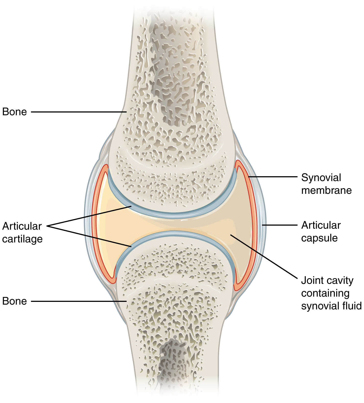
The inner surface of each capsule is lined with a synovial membrane which bounds the synovial cavity in each joint. Movement in one plane Uniaxial movement. Structure of a Synovial Joint - Anatomy Practice.
Flashcards Glossary Test Yourself. B The hinge joint of the elbow works like a door hinge. The structure of a long bone humerus of arm Figure 59.
These types of joints lack a joint cavity and involve bones that are joined together by either hyaline cartilage or fibrocartilage Figure 931. The right elbow joint lateral view Sets found in the same folder. In this article we shall look at the anatomy of the elbow joint.
As the name indicates at a cartilaginous joint the adjacent bones are united by cartilage a tough but somewhat flexible type of connective tissue. The Knee Joint Drag the correct label to the appropriate structure of the knee joint. Structure of a hair and hair follicle Figure 410.
It is classed as a hinge-type synovial joint. The seven bones of the neck are called vertebrae. Learn vocabulary terms and more with flashcards games and other study tools.
The Circulatory Supply to a Mature Bone. Muscles that move the hand and fingers posterior view middle layer Art-labeling Activity. Anatomy and Physiology questions and answers.
The lungs flank the heart. Take the Chapter Practice Test to assess your progress and get your personalized study plan. Endochondral Ossification Jan 12 Thurs Read Chapter 7 of text.
The six types of synovial joints allow the body to move in a variety of ways. Structure of a nail. Bio 165 Chapter 2.
A typical synovial joint Figure 43. Human skull superior view top of cranium removed Figure 511. Structural classification of articulations - Anatomy Practice.
Joint articular Capsule Encloses the joint cavity Consists of 2 parts. The body spinous process of a typical vertebra forms the rounded central portion that faces anteriorly in the human vertebral column. Slipping movements only Nonaxial movement.
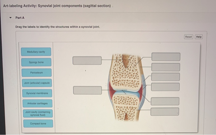
Chapter 6 Osseous Tissue. Human skull anterior view. Start studying The Structure of a Synovial Joint Sagittal section.
After you have studied the bones in lab label the drawings as a self-test. Anatomical structure of the hip joint 1 of 2 Abutabular forum Tobolomical ligament Ligament of head of femur Articular cartilage Articular capsuide Joint cavity llofemoral ligamont Pubic bone Grade trochanter Pued hip bones Femur hotel con anteriori Arterie. The Structure of Osseous Tissue.
The vertebrae articulate with the corresponding ribs. Human skull lateral view. Human skull inferior view mandible removed Figure 512.
A Case Study on Bone Tissue Structure and Repair. Part A Drag the correct label to the appropriate location to identify the types of synovial joints. Muscles that move the hand and fingers posterior view deep layer.
Synovial Joints Anatomy And Physiology
Ch 8 Art Labeling Activity Structure Of A Synovial Joint Anatomy Practice Flashcards Quizlet

Synovial Joints Anatomy And Physiology I
Solved Art Labeling Activity Synovial Joint Components Chegg Com
Comments
Post a Comment